Angiology
Intermediate care unit in the 4th Department of Medicine
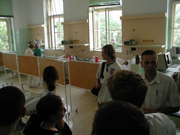
Students are briefly introduced to patients history and are free to ask them questions.
Patients in this ward suffer from poor blood flow in peripheral arteries (excluding cotronsry and cerebral, who are treated in cardiology and neurology, respectively). The major reason for the dissease (ethiopathogenesis) is usually atherosclerosis.
During medical studies, students are supoposed to take patients history, perform elementary physical examination and refere the supervisor about his/her findings.
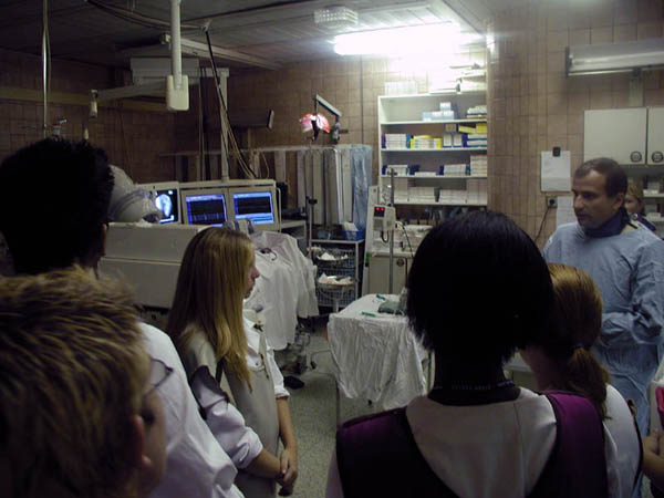
Electrophysiology Lab
2nd Department of Medicine
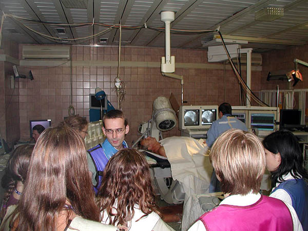
Detailed studies of intracardiac action potential propagation are performed here in order to diagnose and treat various arrhythmias.
The procedure is being explained to the students and details on pathophysiology and treatment of the patient being examined are given.
Students can ask both assistant (blue jacket) and principal investigator (by the monitors).
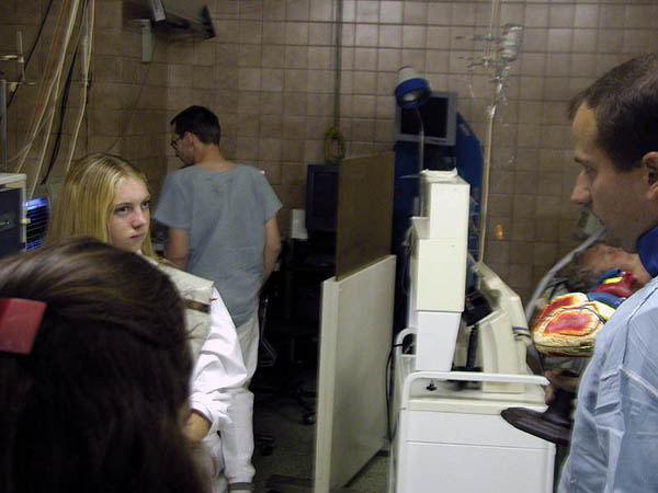
Cath-Lab
2nd Department of Medicine
Coronary angiography are performed here. Most of the patients being examined suffer from disseases related to cardiac ischemia (insufficient blood supply of the myocardium due to obstruction of coronary arteries) such as angina or myocardial infarction.
These diagnoses are among the most common causes of deaths in our population.
Students can see the procedure both on X-ray linked monitor and live through the window with X-ray shielding.
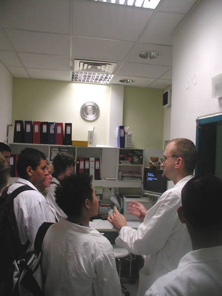
Cardiac Electrophysiology
2nd Department of Medicine
This sophisticated method enables detailed study of endocardial distribution of electrical field of the heart and moiniinvasive treatment of some arrhythmias as ectopic pacemakers or accesory pathways.
The principles of cardiac imaging methods demonstarted on a sophisticated model of the heart (right). The model can be unfolded and used to demonstrate intracardiac parameters.
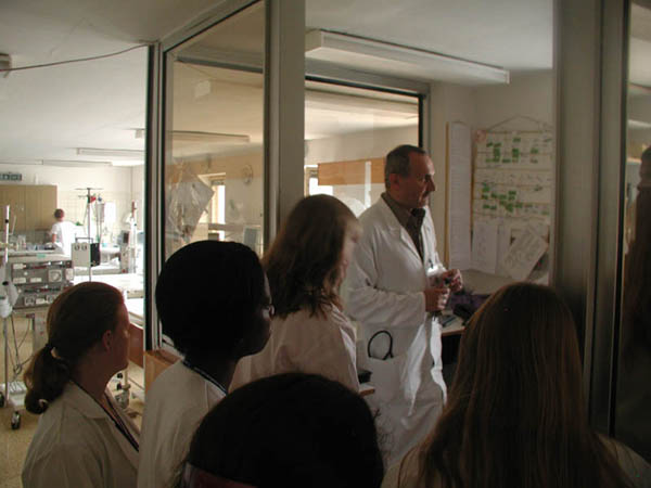
Electrophysiology Lab
2nd Department of Medicine
Operator explains the case after the procedure. Some specific features and events of this particular procedure are discussed, and also expected outcomes and prognosis.
On the monitors behind (left), Intracardiac ECG traces can be seen.
Hemodialysis Unit
1st Department of Medicine
This hemodialysis unit comprises 6 chronical and 2 acute, fully equipped beds. This surprisingly low number is sufficient to cover the needs for dialysis in such a big hospital since the number of renal transplants in our country is one of the highest in Europe.
Differences between acute and chronical hemodialysis are discussed.